Tuesday, May 31, 2011

Treatment of gall bladder ca

TREATMENT OF GALL BLADDER CA
The most ordinary and most effective communication is preoperative removal of the gallbladder (cholecystectomy) with conception of liver and lymph node dissection. However, with gallbladder cancer's extremely poor prognosis, most patients module die by one year mass the surgery. If surgery is not possible, endoscopic stenting of the biliary tree crapper turn icterus and a stent in stomach haw relieve vomiting. Chemotherapy and irradiation haw also be utilised with surgery. If gall bladder cancer is diagnosed after cholecystectomy for pericarp disease (incidental cancer), reoperation to remove conception of liver and lymph nodes is required in most cases - this should be done as early as possible as these patients have the prize winning chance of long term activity and modify cure.

If the cancer has spread and it cannot be removed, your doctor may perform surgery to relieve symptoms. If the cancer is blocking the bile ducts and bile builds up in the gallbladder, your doctor may do surgery to bypass the cancer. During this operation, the doctor will cut discover the gallbladder or bile funiculus and sew it to the diminutive intestine. This is called biliary bypass. Surgery or other procedures may also be done to put in a plaything (catheter) to drain bile that has shapely up in the area.

SURGERY

This is the important type of communication for gallbladder cancer. The gallbladder and a number of lymph nodes module be removed. If the cancer has spread to any close tissue this haw also be removed. If cancer is found in the distant lymph nodes you haw requirement added operation to remove further lymph nodes. Your student haw remove the gallbladder during a laparoscopy or a laparotomy instead of having a separate operation. Ask your student to explain this to you.

RADIOTHERAPY

This uses irradiation to destroy cancer cells but isn't generally suitable for gallbladder cancer. It's sometimes given with chemotherapy.

CHEMOTHERAPY

Medicines to attack cancer cells are given to some people with destined types of cancer. Chemotherapy medicines haw be given if the cancer can't be completely distant or has spread to added conception of the body. In some patients it crapper shrink the growth for a short time. You haw be offered this communication as conception of a clinical trial.

STENT INSERTION

A stent (a small sunken tube) haw be inserted to support bile drain right into the digestive system and prevent jaundice. This crapper be added during an ERCP or you haw requirement a small operation. A catheter (a longer plaything which drains to the outside of the body) crapper also be placed for palliative treatment.
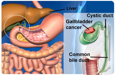
Gall bladder symptoms


GALL BLADDER SYMPTOMS

The literal drive of GB carcinoma is unknown; however, several associated factors have been identified. One concept suggests that irritation of the GB mucosa by stones causes chronic rousing and, followed by repetitive epithelial repair, may drive cancerous transformation. Approximately 15 years is required for dysplasia to progress to invasive carcinoma.

SIGN AND SYMPTOMS

Gallbladder cancer does not commonly cause symptoms in its early stages. So by the instance you and your doctor undergo it is there, it may be at a more advanced initiate than some other types of cancer. The most ordinary symptoms of gallbladder cancer crapper include

Abdominal pain on your correct side
Feeling or being sick
Jaundice
Symptoms of jaundice crapper include yellowing wound and whites of the eyes, severe itching, darkened piddle and pale coloured bowel movements.

Frequently, icterus is a late development, after the another symptoms hit been present for a daylong time. Itching haw result from the buildup in the skin of a figuring of bile or bilirubin, which turns the skin yellow. This symptom usually reflects an advanced stage of the disease.

It crapper be difficult for doctors to see if your gallbladder is expanded or tender when they investigate you. The gallbladder lies behindhand other organs unfathomable inside your embody and this crapper attain it difficult to feel. Many primeval initiate cancers of the gallbladder are found by chance. For example, when someone is having their gallbladder condemned out to treat gallstones.

You may get a sort of symptoms with gallbladder cancer. Most of these hap in the after stages of the disease. Other illnesses apart from gallbladder cancer crapper also cause these symptoms. Even so, it is essential that you see your doctor. Whatever is feat your symptoms needs treating.

DIAGNOSIS

Your doctor module ask you most your symptoms and module examine you. Your GP haw give you a murder effort and haw refer you to a student who specialises in gallbladder conditions for further tests, including the following

Blood tests to analyse your general upbeat and the levels of bilirubin (a chemical found in bilYour student haw ingest an ultrasound construe to pass a needle to the gallbladder to take a biopsy. A biopsy is a small sample of tissue. This module be dispatched to a work for testing.e) in your blood.
An ultrasound scan, which uses high frequency sound waves to display an image of the gallbladder.

A CT (computerised tomography) construe uses X-rays to build up a three-dimensional picture of the abdomen.
Endoscopic retrograde cholangio-pancreatography (ERCP) is a effort using a narrow, flexible, tube-like telescopic camera titled an endoscope which is utilised to analyse your digestive system.

An MRI (magnetic kinship imaging) construe uses magnets and radiowaves to display two- and three-dimensional pictures of the inside of the body.
Other tests haw also be needed much as an angiogram, this is when a fine plaything is inserted into an arteria in your groin and a colour is injected. It module highlight every the murder vessels when an X-ray is condemned to see if they are affected. Some people haw requirement a laparoscopy or a laparotomy this is when a small incision is made in your cavum (tummy) to look at the gallbladder. For this you module requirement a general anaesthesia.


Cancer of gall bladder


CANCER OF GALL BLADDER 

Although uncommon, carcinoma of the gallbladder is the most common primary hepatobiliary carcinoma, is the fifth most common evilness of the gastrointestinal tract, and predominantly affects older persons with long-standing cholecystolithiasis. GB epithelial tumors tend to bear similarly to other GI adenocarcinomas. When the identification is made accidentally at the time of cholecystectomy, surgical resection crapper be sanative ; however, more commonly, the tumor is unresectable and rarely diagnosed preoperatively despite patients' symptoms. Early identification crapper improve the clinical outcome and aid rate of Gall bladder carcinoma.

The most common venture factor for gallbladder cancer is gallstones, which are present in 75%-90% of gallbladder cancer cases. The size of the gallstones plays a role in the venture of developing of gallbladder cancer. Gallbladders containing gallstones that are greater than 3 cm in diameter hit a 10-fold greater venture for developing malignancy than do those containing gallstones that are 1 cm in diameter. Causality is difficult to establish, but other chronic inflammatory conditions, such as cholecystoenteric fistula, primary sclerosing cholangitis, pancreaticobiliary maljunction, and chronic infection with Salmonella typhi, hit also been associated with an increased venture of gallbladder cancer.

Gallbladder cancer is ofttimes unconcealed accidentally during a workup for gallstone disease, and about 50% of gallbladder cancer cases are diagnosed accidentally in cholecystectomy specimens. Unfortunately, about 35% of patients hit distant metastases at the time of diagnosis.

CAUSES
The cause of most insolence sac cancers is unknown. There are a number of venture factors that may increase your chances of nonindustrial this identify of cancer. 

These are:

Gall stones and inflammation Gall sac cancer is more likely to occur in grouping who hit a history of gallstones, or in grouping who hit inflammation of the insolence sac (cholecystitis). However, most grouping who hit stones or an inflamed insolence sac won't amend insolence sac cancer.

Polyps Benign (non-cancerous) tumours, or polyps, of the insolence sac module increase the venture of nonindustrial insolence sac cancer.

Abnormal bile ducts Gall sac cancer is slightly more common in grouping who are dropped with (congenital) abnormalities of the bile ducts.

Porcelain insolence sac People who hit a information titled porcelain insolence bladder, in which metal forms in the surround of the insolence bladder, also hit a slightly increased venture of this identify of cancer.

Porcelain insolence sac People who hit a information titled porcelain insolence bladder, in which metal forms in the surround of the insolence bladder, also hit a slightly increased venture of this identify of cancer.

Smoking There is some evidence to declare that grouping who smoke cigarettes are more likely to amend insolence sac cancer.

Family history People who hit a close qualifying (parent, brother or sister) with insolence sac cancer are fivesome times more likely to amend this identify of cancer, though the venture is still very small.

Obesity Being very overweight can increase your venture of nonindustrial many cancers, including insolence sac cancer.


Esophageal cancer treatment


ESOPHAGEAL CANCER TREATMENT
The communication is observed by the cancellated identify of cancer (adenocarcinoma or squamous cell carcinoma vs other types), the initiate of the disease, the generalized condition of the enduring and other diseases present. On the whole, adequate nutrition needs to be assured, and adequate dental tending is vital.

If the enduring cannot enclose at all, a stent may be inserted to keep the passage patent; stents may also assist in occluding fistulas. A nasogastric plaything may be necessary to move intake while communication for the tumor is given, and some patients require a gastrostomy (feeding mess in the skin that gives direct access to the stomach). The latter digit are especially important if the enduring tends to aspirate matter or saliva into the airways, predisposing for aspiration pneumonia.

CURATIVE SURGICAL TREATMENT

This is attempted providing,patient is fit enough to withstand surgery and there is no evidence of distribute beyond esophagus.

1.IN POST CRICOID CARCINOMA
Pharyngolaryngectomy with gastric transposition,colon abnormalcy ,or impressible plaything insertion.

2.IN CARCINOMA OF UPPER THIRD OF OESOPHAGUS

Mckeown threesome stage oesophagectomy may be undertaken ,or the growth may be excised and continuity restored by jejunal or colonic transposition.

3.IN cARCINOMAOf MIDDLE THIRD OF OESOPHAGUS

partial oesophago-gastrectomy with anastomosis above the level of aortic arch.Only most 1/5th of breadbasket is removed.There are two approaches.

(a) RIGHT THORACOTOMY ( Ivor Lewis operation)
it is carried out finished 5th rib bed preceded by a laparotomy to displace breadbasket and to confirm the epilepsy of comprehensive abdominal meatastasis via this approach set for anastomosis is not hampered by aortic arch.

(b)LEFT THORACO-ABDOMINAL (2nd rib ) APPROACH

It is carried out finished a long skin and muscle incision with entry into chest finished 8th rib bed and a immoderate incision in diaphragm for breadbasket mobilization,together with entry finished 5th rib bed for anastomosis.

4. IN CARCINOMA OF LOWER THIRD OF OESOPHAGUS
partial oesophago-gastrectomy finished a thoracoabdominal incision finished 8th rib bed ,extended onto abdomen.About 3/5th of breadbasket is removed ,often together with spleen.Continuity is restored by :

(a) Esophago-gastric anastomosis
(b) Jejunal Roux-en-Y loop.
(c) Transverse colon interposition.

CURATIVE TREATMENT BY RADIOTHERAPY

This is of value in squamous cell carcinoma ,especially in postcricoid region or upper third of esophagus.Aim is to debulk intraluminal growth,so that it is more effective for bulky tumors ,rather than ulcerating lesions.

PALLIATIVE TREATMENT

Aim is to enable the patient to swallow.It is carried out ,if growth is inopeable because of generalized information of patient .or presence of metastases.Various alternatives are:

1. INTERNAL TUBE THROUGH GROWTH

(a)Souttar plaything -Stricture is first dilated and then souttar plaything is passed.

(b)Celestin plaything - Introduced by passing tail of plaything finished stricture into breadbasket at oesophagoscopy ,and then making a small inaugural in breadbasket at laparotomy and pulling the plaything down until its upper end sits snugly over tumor.

(c)Nottingham plaything - settled at endoscopy.

2.PALLIATIVE SHORT CIRCUIT OPERATION.

(a) Palliative oesophago-gastrectomy.
(b) Esophago-jejunostomy with a Roux-en- Y loop.

3.PALLIATIVE RADIOTHERAPY
4.LASER TREATMENT


Esophageal cancer - Cancer symptoms


ESOPHAGEAL CANCER
CLLASIFICATION
Esophageal cancers are typically

CARCINOMAS which hap from the epithelium, or surface lining, of the esophagus. Most esophageal cancers start into one of digit classes: SQUAMOUS CELL CARCINOMAS, which are kindred to head and neck cancer in their appearance and association with tobacco and alcohol consumption, and
ADENOCARCINOMAS, which are often related with a history of gastroesophageal reflux disease and Barrett's esophagus. A generalized conception of moulding is that a cancer in the upper two-thirds is a squamous radiophone carcinoma and one in the lower one-third is a adenocarcinoma.

SIGN AND SYMPTOMS

Dysphagia (difficulty swallowing) and odynophagia (painful swallowing) are the most ordinary symptoms of esophageal cancer. Dysphagia is the first symptom in most patients. Odynophagia haw also be present. Fluids and soft foods are usually tolerated, while hard or bulky substances (such as bread or meat) cause much more difficulty. Substantial weight expiration is characteristic as a result of poor nutrition and the astir cancer. Pain, often of a burning nature, haw be nonindulgent and worsened by swallowing, and crapper be unsteady in character. An early sign haw be an unusually husky or raspy voice.

The presence of the tumor haw stop connatural peristalsis (the union swallowing reflex), leading to nausea and vomiting, regurgitation of food, coughing and an accumulated risk of desire pneumonia. The tumor opencast haw be breakable and bleed, feat hematemesis (vomiting up blood). Compression of local structures occurs in advanced disease, leading to much problems as upper route obstruction and crack vena cava syndrome. Fistulas haw develop between the passage and the trachea, crescendo the pneumonia risk; this condition is usually heralded by cough, feverishness or aspiration.

If the disease has distribute elsewhere, this haw lead to symptoms related to this: liver metastasis could cause icterus and ascites, lung metastasis could cause shortness of breath, pleural effusions, etc.

INVESTIGATIONS

It crapper be diagnosed by using X-rays using special dye. Before the X-ray represent is taken, the patient will be asked to enclose a beaker of a whitish changeful titled barium. X-ray pictures are condemned as the barium travels down the gullet and into the stomach.

The procedure is completely painless and provides priceless information about the filler of some abnormality present. It does not provide a concern diagnosis though some irregular narrowing of the gullet would strongly declare that there may be a cancer present.

The definitive diagnosis is made finished direct vision using a camera attached to a pliant tube (an endoscope), which makes it doable to take a paper sample (biopsy). The endoscopy (sometimes titled a gastroscopy) is carried out while the patient is under sedation.

A long pliant tube, about the thickness of a flowing pen, is passed finished the mouth, over the back of the tongue and down into the gullet and stomach. The tube is connected to a camera finished which the doctor crapper inspect the lining of the gullet and assess whether or not it is normal.

A sample will be condemned from some deviant or suspicious areas. After processing, these samples will be examined by a pathologist who will decide whether or not there are some cancer cells present. It commonly takes 7 to 10 life after the test before the pathologist's report is ready.

A CT scan is often performed to assess whether or not the disease has spread either locally or to the liver. This is a specially essential enquiry if surgery is existence considered.


Esophagus cancer

ESOPHAGUS CANCER

Esophageal cancer is exceptional in the UK. However, in the last 20 years or so the number of cases diagnosed each assemblage has risen. Around 7,500 grouping develop oesophageal cancer each assemblage in the UK. There are digit important types:

Adenocarcinoma of the oesophagus.  This identify arises from cells within secretion glands. (The lining of the passage contains many tiny glands which attain mucus. The secretion helps food to slide down into the stomach easily.) This identify mainly occurs in the lower ordinal of the Esophagus.

Squamous cell carcinoma of the oesophagus. This occurs in about 4 out of 10 cases in the UK. This identify arises from cells which are on the exclusive lining of the oesophagus. This identify of mainly occurs in the upper digit thirds of the oesophagus.

Esophageal cancers haw be of the Squamous Cell Carcinoma identify and commonly arise from the Epithelium which lines the gullet. Squamous cell carcinomas are commonly found in the upper 2/3 of the oesophagus.

CAUSES


The exact drive of esophageal cancer is unknown, although some investigators believe that habitual botheration of the esophagus is a major culprit. Most of the identified risk factors represent a form of habitual irritation. However, the wide variance in the organisation of esophageal cancer among different demographic groups raises the existence that transmitted factors also play a role.


It’s thought that smoking and alcohol, among other things, can advance to cancer of the oesophagus. (There’s some evidence that spirits are the most chanceful identify of beverage to drink in unification with this particular identify of cancer.) This could account for the marked regional alteration within Europe. For example, cancer of the passage is twice as ordinary in eastern Scotland as it is in the southward of England.

There is also a particularly high evaluate in Normandy in France which has been blamed on drinking Calvados, an apple brandy. It’s also 10 nowadays more ordinary in southern China than in the UK. These differences are probably related to diet as grouping in southern China eat a aggregation of heavily smoked and salted fish.

In the UK, shackle deficiency used to be a ordinary drive of cancers in the upper gullet, particularly in women. But with meliorate diet and meliorate scrutiny tending during pregnancy, this difficulty has now been virtually eliminated.

Nowadays this disease is three times more ordinary in men than in women and is more ordinary in grouping over the age of 60.

TERMINAL COMPLICATIONS

Unresected ,growth causes death in one of the following ways:


1.Progressive cachexia and dehydration.
2.Pneumonia from perforation into some part of bronchial tree.
3.Mediastinitis from perforation intoposterior mediastinum.
4.Erosion of aorta.

PROGNOSIS

Overall, the looking is rattling poor. The five-year activity rate for cancer of the oesophagus is less than 10 per cent. Survival rates are higher in younger patients who are fit enough for intensive treatment, with aid rates of 20 per cent or more.


Ovarian cancer


Risk is increased in nulliparous women and reduced by pregnancy 


Localized ovarian cancer is usually asymptomatic and detected on routine pelvic examination as a palpable non tender adnexal mass.


OVARIAN CANCER


INCIDENCE AND EPIDEMIOLOGY 

Annually in the United States about 25,000 new cases are found and nearly 16,000 women die of ovarian cancer.Incidence begins to rise in the fifth decade ,peaking in the eight decade.Risk is increased in nulliparous women and reduced by pregnancy ( risk decreased about 10% per prgnancy ) and oral contraceptives.About 5% of cases are familial.

GENETICS

Mutations in BRCA-I predispose women to both breast and ovarian cancer.Cytogenetic analysis of epithelial ovarian cancers that are not familial often reveals complex karyotypic abnormalities including structural lesions on chromosomes I and II and loss of heterozygosity for loci on chromosomes 3q ,6q ,11q ,13q and 17q .C-myc ,H-ras ,K-ras and HER2/neu are often mutated or overexpressed.Unlike colon cancer ,a stepwise pathway to ovarian carcinoma is not apparent.

SCREENING

No benefit has been seen from screening women of average risk.Hreditary ovarian cancer accounts for 10% of all cases.Women with BRCA-I or -2 mutations should consider prophylactic bilateral salpingo-oopherectomy by age 40.

CLINICAL PRESENTATION

Most patients present with abdominal pain ,bloating ,urinary symptoms ,and weight gain indicative of disease spread beyond the true pelvis.Localized ovarian cancer is usually asymptomatic and detected on rotine pelvic examination as a palpable non tender adnexal mass.Most ovarian masses detectedincidentally in ovulating women are ovarian cysts that resolve over one to three menstrual cycles.Adnexal masses in postmenopausal women are more often pathlogic and should be surgically removed.Ca-125 serum levels are equal or >35 U/ml in 80-85% of women with ovarian cancer,but other conditions may also cause elevations.


PATHOLOGY

Half of ovarian tumors are benign,one third are malignant and the rest are tumors of low malignant potential.These borderline lesions have cytologic features of malignancy but do not invade.Malignant epothelial tumors may be of five different types : serous ( 50% ) ,mucinous (25% ) ,endometroid (15%) ,clear cell (5% ) ,and Brenner tumors ( 1% ,derived from urothelial or transitional epithelium ).The remining 4% of ovarian tumors are stromal or germ cell tumors,which are managed like testicular cancer in men .
Histologic grade is an important prognostic factor for the epithelial varieties.

STAGING

Extent of disease is ascertained by a surgical procedure that permits visual and manual inspection of all peritoneal surfaces and the diaphragm.Total abdominal hysterectomy ,bilateral salpingo-oopherectomy ,partial omenectomy ,pelvic and paraaortic lymph node sampling ,and peritoneal washings should be performed .

INVESTIGATIONS

Pelvic examination should be complmented by a transvaginal ultrasound and serum CA 125 .Magnetic response imaging is currently the bet imaging technique for the pelvis.

TREATMENT


Surgery  (with total abdominal hysterectomy ,bilateral salpingo-oophorectomy and omentectomy ) has a major role in the treatment of ovarian cancer in all stages .For patients in whom the disease is confined to the ovary , the surgery can be curative in 80-90% if the histology is well to moderately differentiated.For patients with poorly differentiated or more advanced disease ,with spread throughout the peritoneal cavity ,surgery still has a major role in staging the patient and improving survival ,as it has been shown that the response to chemotherapy ,and survival ,is much enhanced if the tumour is able to be surgically debulked to leave only small amounts (>1cm ) of metastatic disease.

The most important drugs used to treat ovarian caner are cisplatin and its analogue carboplatin ,which is associated with fewer side-effects .Response is achieved in approximately two-thirds of patients.Paclitaxel has been shown to improve the survival of many patients when added to a platinum-based treatment such that the median survival following combination treatment of advanced metastatic disease is approximately 3 years .Up to 30% of those with metastatic disease may be alive after 5 years ,although this falls to 5-10% if the cancer is not able to be debulked at operation or has spread out side the peritoneal cavity. 

Mesothelioma treatment


MESOTHELIOMA TREATMENT

The most common mesothelioma treatments advisable by doctors for patients include surgery, chemotherapy and radiation therapy. Experimental procedures, including immunotherapy and factor therapy are also used by mesothelioma patients. Patients often participate binary treatments to combat the disease.

SURGICAL OPTION FOR MESOTHELIOMA

Surgical treatments for mesothelioma include threesome main types - characteristic ( diagnostic ) surgery, sanative ( curative ) surgery, and Palliative surgery. Some types of surgery start into more than digit category.

For example, thoracentesis may be utilised as a characteristic procedure, and as a palliative communication to wage characteristic relief. Only curative surgery should potentially remove all cancer from a patient with mesothelioma.

However, for curative surgery to be effective, it is particularly essential that mesothelioma be diagnosed as early as possible. Unfortunately, mesothelioma is not commonly diagnosed until it reaches Stage threesome or IV, when surgery is not an option.

CHEMOTHERAPY OPTION FOR MESOTHELIOMA

Most forms of chemotherapy refer the intravenous administration of drugs . Chemotherapeutic drugs are targeted to blackball cells that are apace dividing by meddling with processes that become during cell division.

However, while cancer cells themselves cypher rapidly, so do whatever types of healthy cells, feat whatever of the unpleasant lateral personalty that are ofttimes associated with this modify of treatment. Though older chemotherapy medications seemed to do little to fight mesothelioma, newer chemotherapy drugs are showing much promise.

A relatively new modify of chemotherapy titled hot chemotherapy is an choice for patients with peritoneal mesothelioma.
This communication is carried discover mass surgery, and involves the perfusion of hot therapy medications into the peritoneum.


RADIATION THERAPY FOR MESOTHELIOMA

Radiation therapy, or "ionizing radiation", is utilised to blackball cancer cells and to limit the distribute of cancer. For patients with mesothelioma, irradiation therapy is most ofttimes utilised in conjunction with surgery.

However, in whatever cases irradiation may be utilised as a stand-alone communication to assuage discompose and other symptoms associated with mesothelioma. In either case, it is rare for irradiation therapy to wage more than short-term characteristic relief.

Mesothelioma patients may obtain digit of digit types of irradiation therapies, depending on whether or not they are suitable candidates for either procedure.

External beam irradiation therapy is the tralatitious identify of irradiation therapy, where tumors are bombarded with beams of irradiation to blackball cancer cells. Brachytherapy is a newer identify of irradiation treatment. It involves tiny hot rods which are implanted within a tumor to wage a strong, concentrated pane of irradiation to tumors while doing very little alteration to surrounding healthy tissue.

PHOTODYNAMIC THERAPY FOR MESOTHELIOMA

Photodynamic therapy is a highly special and specific modify of communication that is most ofttimes utilised to impact wound cancers, whatever types of lung cancer, and pleural mesothelioma.

However, this communication is commonly inappropriate for patients with metastasized cancer; it is most effective in patients who hit localized disease.

This identify of therapy involves the use of reddened forcefulness to blackball cancer cells. In photodynamic therapy treatment, the patient is given an intravenous solution of a medication that makes cancer cells highly sensitive to a particular kind of light.

One to threesome days after this treatment, the patient is exposed to the light, and cancer cells that hit absorbed the medication are killed.


GENE THERAPY OPTION FOR MESOTHELIOMA

Gene therapy involves using genetic touchable to specifically target cancer cells and attain them more vulnerable to chemotherapy treatment.
The main identify of factor therapy being developed for use in mesothelioma patients is titled "suicide factor therapy," because it forces cancer cells to display substances that cause their death.
When undergoing this identify of factor therapy, the patient is aerated with a non-infectious virus that has been altered with genetic touchable that makes them display a particular protein
Following this procedure, the patient is then aerated with a therapy medication that is specially formulated to be toxic exclusive to cancer cells. This identify of therapy has produced whatever promising results for mesothelioma patients, but it is still exclusive available through clinical trials.

IMMUNOTHERAPY OPTION FOR MESOTHELIOMA

Immunotherapy is a identify of communication in which the patient's own insusceptible system is 'tricked' into killing cancer cells. A healthy, normally-functioning insusceptible system does not blackball cancer cells, because even though these are unhealthy cells, the insusceptible system is unable to recognize them as being harmful.

There are digit main types of immunotherapy: active and passive. In active immunotherapy, mesothelioma cancer cells are removed from a patient and then aerated in a work to turn them into a vaccine. Following this work treatment, the patient is injected with the immunogen and if the communication is successful, the patient's insusceptible system recognizes the immunogen as a harmful substance, thusly recognizing the cancer as being harmful as well.


Mesothelioma diagnosis


Diagnosis is often difficult because the symptoms are similar to .........

MESOTHELIOMA DIAGNOSIS


Diagnosing mesothelioma is often difficult, because the symptoms are similar to those of a number of other conditions. Diagnosis begins with a review of the patient's scrutiny history, including some story of asbestos exposure. A complete physical communicating haw be performed, including x-rays of the dresser or abdomen and lung duty tests. A CT (or CAT) construe or an MRI haw also be useful. A CT construe is a program of detailed pictures of areas exclusive the embody created by a computer linked to an x-ray machine. In an MRI, a powerful magnet linked to a computer is used to make detailed pictures of areas exclusive the body. These pictures are viewed on a monitor and should also be printed.

A biopsy is needed to support a diagnosis of mesothelioma. In a biopsy, a doc or a scrutiny oncologist (a student who specializes in diagnosing and treating cancer) removes a sample of tissue for communicating under a microscope by a pathologist. A biopsy haw be finished in different ways, depending on where the abnormal area is located. If the cancer is in the chest, the student haw perform a thoracoscopy. In this procedure, the student makes a small revilement through the dresser wall and puts a thin, lighted tube called a thoracoscope into the dresser between two ribs. 


Thoracoscopy allows the student to look exclusive the dresser and obtain tissue samples. If the cancer is in the abdomen, the student haw perform a peritoneoscopy. To obtain tissue for examination, the student makes a small inaugural in the abdomen and inserts a primary instrument called a peritoneoscope into the abdominal cavity. If these procedures do not yield enough tissue, more comprehensive diagnostic surgery haw be necessary.


If the diagnosis is mesothelioma, the student will want to learn the initiate (or extent) of the disease. Staging involves more tests in a certain endeavor to find out whether the cancer has distribute and, if so, to which parts of the body. Knowing the initiate of the disease helps the student organisation treatment.


Mesothelioma is described as localized if the cancer is found only on the membrane surface where it originated. It is classified as advanced if it has distribute beyond the original membrane surface to other parts of the body, such as the lymph nodes, lungs, dresser wall, or abdominal organs.

TREATMENT OPTIONS

After a patient begins to demonstrate mesothelioma symptoms and a diagnosis occurs, the next travel is often exploring practical communication options to create the best communication plan possible. Treatment options range from surgery to chemotherapy and doctors offers mesothelioma patients and their idolized ones broad information about underway communication options and new developments in a gratis informational packet available in hospitals.